Phenotyping regenerants in plant tissue culture
Rising plants from tissue culture is a frequent application in research, plant propagation, plant breeding, and genetic engineering. Tissue cultures can regenerate to fully functional plants. When plants start growing on nutrient-supplied artificial media, first stages do not yet have chloroplasts, but as soon as light exposure starts, newly grown tissue develops chloroplasts and starts photosynthesizing. To figure out whether cultivation is successful, constant monitoring of the regenerating plants.
Beyond visual inspection, numerical data on growth and development are highly desirable, and an automated sample evaluation makes the process faster, standardized, and documented. At this point, LemnaTec’s phenotyping technology delivers high quality images and advanced, AI-supported image processing. When taking images of the samples, phenotypic traits are instantly derived by the LemnaTec software.
The analytical software separates the plants from the background and recognizes the plants as single objects. All detected objects can be measured for various factors.
Regenerant plants usually grow on dishes, and after imaging, the analysis aims to segment target objects and measure their properties. For such a dish with a group of plants on growth medium, key steps in the analysis process are: first, locating the plants, second, color segmenting the image, third object identification, and fourth, determining and storing geometrical parameters of the objects.
After this process, results are not only given as visualization, but also as numerical data table that can be used for data visualization and statistics.
As result of the analysis, plant 3 has the highest fraction of green tissue, in plants 2 and 4 substantial areas of pale tissue are visible, and in plant 1 more than half of the plant is pale. Of course, it depends on the context of the cultivation how to interpret such fractions of tissue colors. LemnaTec’s image processing tools can provide application-specific data and via machine learning, the segmentation can be trained to sample-specific traits.

The LemnaGrid software has a graphical user interface where devices, i.e., pre-defined functions in image processing, can be combined to image processing workflows. These functions enable stepwise analyzing images with the aim to segment target objects and measure their properties.
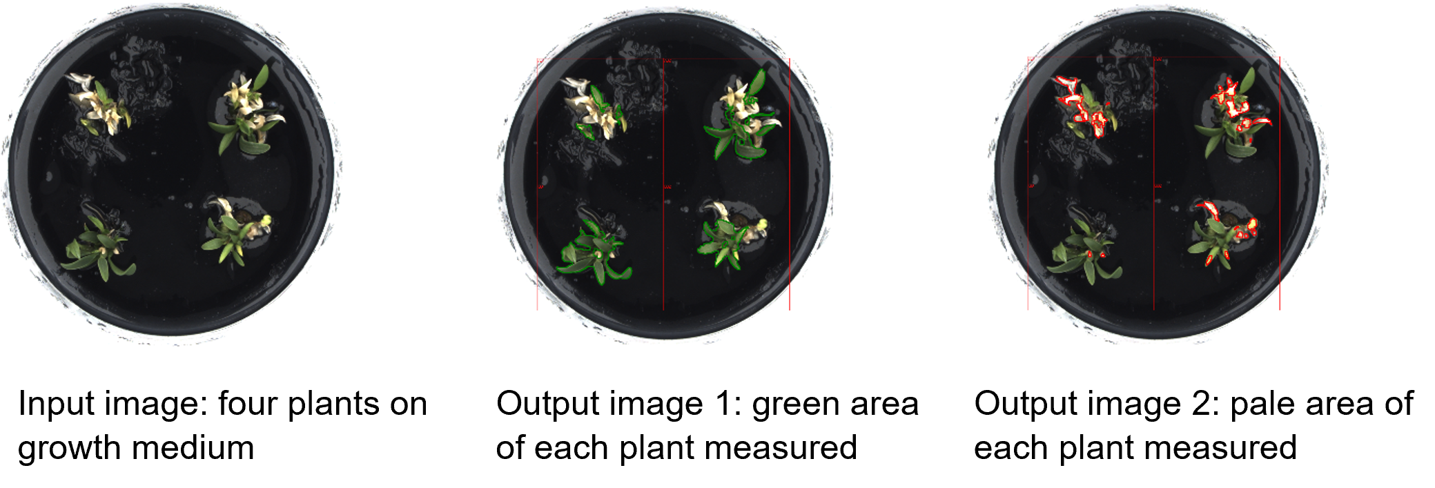
In the current example, four regenerating plants on a plate with growth medium were imaged with a LemnaTec PhenoAIxpert. The software analyzed the image and determined green and pale areas of all four plants. The green areas refer to tissues that have already built up chloroplasts, while the pale parts of the plants are those that have grown chloroplast-free on the nutrient-supplied medium. Fractions of both tissue types can be determined from the image.

Analysis of green and pale leaf area for four regenerant plants on a plate.