Fluorescence imaging in physiological phenotyping
Fluorescence provides access to pigment-related data that are not provided by visible light images. Most prominently, chlorophyll emits fluorescence after excitation by light, but other pigments and breakdown products of chlorophyll can emit fluorescence, too. Chlorophyll causes red fluorescence that dominates the light emission due to high abundance of chlorophyll. The appearance of yellowish fluorescence hints at reduced chlorophyll content, or even absence of chlorophyll, as this signal is caused by breakdown products of chlorophyll or background pigments. Therefore, fluorescence imaging can be used as indicator for stress and senescence, with stressed or senescing tissues appearing yellow while intact tissue appears red.
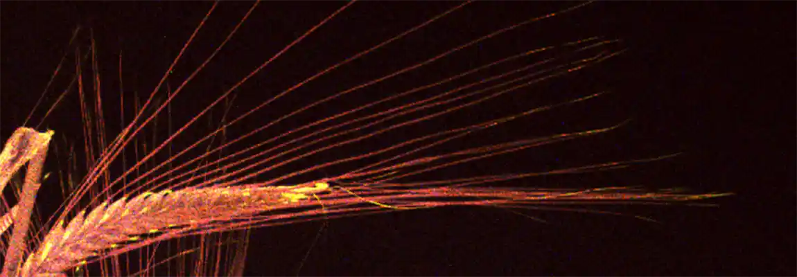
With LemnaTec’s innovative imaging technologies, fluorescence analyses are enabled at whole-plant level. Using high resolution cameras, it is possible to analyze fluorescence signals in details, for example for awns and kernel hulls of cereal inflorescences. Imaging red and yellow fluorescence concomitantly in one image, analyses of stress and senescence are facilitated.
The plant in the example images was kindly provided by IPK Gatersleben, Germany, and images were recorded in the LemnaTec phenotyping system at the IPK.

Overlay of RGB and Fluorescence images acquired with a LemnaTec PhenoAIxpert HT

Detailed view on fluorescence of awns and spikes

Fresh and senescent leaves differ in fluorescence
